Facial tumours of Tasmanian Devils are among the extremely rare cases of transmissible cancer and threaten the survival of this species. They are extremely interesting for medical research because they can be used to unravel new cancer molecular mechanisms and their interplay with the immune system. Scientists at the Center for Molecular Medicine (CeMM), the Vetmeduni Vienna and the MedUni Vienna have succeeded in using modern analysis methods to elucidate key molecular mechanisms that are crucial for the transferability of these cancer cells.

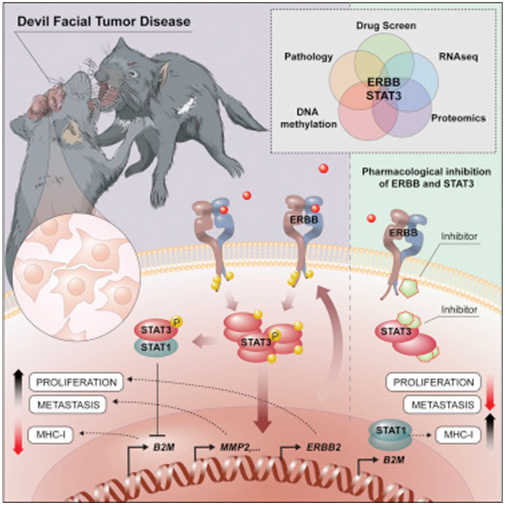
Tumours can usually only proliferate in the organism from which they originated. This is due to a large number of molecular security mechanisms with which the immune system repels and destroys foreign tissue. An exception to this is the Tasmanian Devil, the largest living carnivorous marsupial in the world: a deadly facial tumour has been spreading at a rapid pace for over two decades in this species.

How these cells escape the immune system of its recipient has long been a mystery. Now, the groups of Andreas Bergthaler (CeMM) and Richard Moriggl, head of the Ludwig Boltzmann Institute for Cancer Research and Professor of Functional Cancer Genomics at the Vetmeduni and MedUni Vienna,

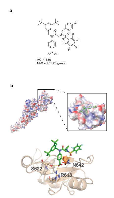
found that certain receptor molecules on the surface of the cancer cells, ERBB receptors, have unusually high activity. The activation of these receptors influences the cell's epigenetic make-up via STAT3. As a result, the number of molecules that the immune system uses to recognize the cell is reduced, while cell division accelerates and factors for the metastasis of the tumour cells are increasingly produced. Furthermore, inhibiting the ERBB receptor with a specific drug can kill cancer cells in a targeted manner. This could play an important role in treating this communicable tumour before the Tasmanian Devil is completely eradicated. In addition, the basic biological principles of invasion and fixation in new tissues are of crucial importance, even in the case of non-communicable tumours, in particular cancer metastases. A better molecular understanding of these relatively exotic communicable tumours can provide important insights into the basic biological mechanisms of cancer.

Published in Cancer Cell

Lindsay Kosack, Bettina Wingelhofer, Alexandra Popa, Anna Orlova, Benedikt Agerer, Bojan Vilagos, Peter Majek, Katja Parapatics, Alexander Lercher, Anna Ringler, Johanna Klughammer, Mark Smyth, Kseniya Khamina, Hatoon Baazim, Elvin D. de Araujo, David A. Rosa, Jisung Park, Gary Tin, Siawash Ahmar, Patrick T. Gunning, Christoph Bock, Hannah V. Siddle, Gregory M. Woods, Stefan Kubicek, Elisabeth P. Murchison, Keiryn L. Bennett, Richard Moriggl and Andreas Bergthaler

Doi: https://doi.org/10.1016/j.ccell.2018.11.018

The Janus kinase-signal transducers and activators of transcription (JAK-STAT) signaling pathway is critical in tuning immune responses and its dysregulation is tightly associated with cancer and immune disorders. Disruption of interleukin (IL)-15/STAT5 signaling pathway due to the loss of IL-15 receptor chains, JAK3 or STAT5 leads to immune deficiencies with natural killer (NK) cell abnormalities. JAK1, together with JAK3 transmits signals downstream of IL-15, but the exact contribution of JAK1 to NK cell biology remains to be elucidated. In this study we show that deletion of NK cell-intrinsic JAK1 leads to an almost complete loss of NK cells in the spleen, blood, and liver, proving a crucial role of JAK1 in peripheral NK cells. The absence of one allele of Jak1 suffices to drastically impair NK cell function whereas the deletion of JAK2 in NK cells has no impact on their survival or maturation. We thus propose that in contrast to currently used JAK1/JAK2 inhibitors, the use of JAK2-specific inhibitors would be advantageous for the cancer patients by leaving NK cells intact.

Publication in Frontiers in Immunology

Agnieszka Witalisz-Siepracka, Klara Klein, Daniela Prinz, Nicoletta Leidenfrost, Gernot Schabbauer, Alexander Dohnal and Veronika Sexl

Loss of JAK1 Drives Innate Immune Deficiency (2019), doi: 10.3389/fimmu.2018.03108

See also Video:  https://drive.google.com/open?id=1lUNFRP4aLRZxr4-9VQkKV30IwV2EppKh

STAT1 exists as two alternatively spliced isoforms, STAT1α and STAT1β; the latter lacks the C-terminal transactivation domain (TAD). Our previous study with gene-modified mice expressing only the STAT1β isoform (Stat1β/β) demonstrated that STAT1β is capable of inducing a subset of IFNγ-responsive genes but the reason for the gene-selectivity remained unclear. In this study we used primary macrophages form wild-type and Stat1β/β mice to characterize the role of the C-terminal TAD in the transactivation and cofactor recruitment to paradigmatic IFNγ-responsive genes. Our key discoveries are that the STAT1β isoform is differentially required for (i) the recruitment of the Mediator coactivator complex and the transition of poised RNA polymerase II (Pol II) into productive elongation, (ii) the association of the general transcription factors TFIIH and p-TEFb to promoter elements specifically at late time points after stimulation or (iii) the establishment of active histone marks and the recruitment of Pol II to the STAT1 and IRF1 co-regulated gene promoters.

Collectively, our results shed new light on the communication of STAT1 with the transcriptional machinery and provide mechanistic insights into isoform-specific transcriptional activities of STAT1.

Publication in Frontiers in Immunology

Matthias Parrini, Katrin Meissl, Mojoyinola Joanna Ola, Therese Lederer, Ana Puga, Sebastian Wienerroither, Pavel Kovarik, Thomas Decker, Mathias Müller  and Birgit Strobl

The C-Terminal Transactivation Domain of STAT1 Has a Gene-Specific Role in Transativation and Cofactor Recruitment (2018), doi: 10.3389/fimmu.2018.02879

RNA helicase DDX3X is important for many aspects of RNA metabolism and RNA translation. In addition, several publications have highlighted a role for DDX3X in immunity, as it contributes to the induction of type I IFNs. However, the in vivo relevance of DDX3X in cells of hematopoietic origin remains unexplored. Using mouse genetics, we demonstrate that DDX3X makes important contributions to innate immunity against pathogens beyond its role in IFN induction, by influencing hematopoiesis as well as the transcription of many antimicrobial genes. By comparison between cells from male or female animals we show that DDX3X functions are in part compensated by its Y-chromosomal homologue DDX3Y. This suggests that DDX3X may be one of the factors contributing to well-established differences of the male and female immune systems. 

Publication in PLoS Pathogens

Daniel Szappanos, Roland Tschismarov, Thomas Perlot, Sandra Westermayer, Katrin Fischer, Ekaterini Platanitis, Fabian Kallinger, Maria Novatchkova, Caroline Lassnig, Mathias Müller, Veronika Sexl, Keiryn L. Bennett, Michelle Foong-Sobis, Josef M. Penninger and Thomas Decker

The RNA helicase DDX3X is an essential mediator of innate antimicrobial immunity (2018), https://doi.org/10.1371/journal.ppat.1007397

Anaplastic Large Cell Lymphomas (ALCL) is a rare type of lymphoma comprising approximately 16 percent of all T cell lymphomas. The molecular analysis of human tumour samples to find new, personalised therapeutic targets and their validation in tumour models has become a attractive approach in cancer research, which can advance clinical management of cancer patients. This is even more important for rare tumours, like ALCL, in which researchers rely on a very small number of patients. New research led by the international ERIA consortium and with the participation of the SFB members Richard Moriggl, Mathias Müller, Birgit Strobl has now identified that all sub-types of ALCL rely on the same signalling pathway for survival.

TYK2 is a member of the JAK family of tyrosine kinases that is involved in chromosomal translocation-induced fusion proteins found in some anaplastic large cell lymphomas (ALCL). Here the authors show that TYK2 is highly expressed in all cases of human ALCL and it prevents apoptotic cell death in ALCL human cell lines by increasing the expression of Mcl1, a pro-survival member of the BCL2 family. Moreover, TYK2 inhibitors are able to induce apoptosis in ALCL cells. Therefore, TYK2 represents an attractive drug target due to its unique enzymatic domain and TYK2-specific inhibitors show promise as novel targeted inhibitors for ALCL. This is an urging therapy that still needs to be developed to treat immunological disorders, such as rare lymphomas.

Publication in Leukemia
Nicole Prutsch, Elisabeth Gurnhofer, Tobias Suske, Huan Chang Liang, Michaela Schlederer, Simone Roos, Lawren C. Wu, Ingrid Simonitsch-Klupp, Andrea Alvarez-Hernandez, Christoph Kornauth, Dario A. Leone, Jasmin Svinka, Robert Eferl, Tanja Limberger, Astrid Aufinger, Nitesh Shirsath, Peter Wolf, Thomas Hielscher, Fritz Aberger, Johannes Schmoellerl, Dagmar Stoiber, Birgit Strobl, Ulrich Jäger, Philipp B. Staber, Florian Grebien, Richard Moriggl, Mathias Müller, Giorgio G. Inghirami, Takaomi Sanda, A. Thomas Look, Suzanne D. Turner, Lukas Kenner and Olaf Merkel

Doi: https://doi.org/10.1038/s41375-018-0239-1

Hematopoietic stem cells (HSCs) sustain blood and immune cells in the body and are therefore crucial for our survival. HSCs are at rest, but as soon as blood needs to be formed - like after blood loss or chemotherapy - they are quickly activated to compensate for this loss. After completing their mission, they must return a dormant state. A team of scientists, including Christoph Bock (CeMM) and Veronika Sexl (Vetmeduni Vienna) – lead by Manuela Baccarini (Max F. Perutz Laboratories (MFPL)) – has now shown how intracellular signal transmissions can maintain this delicate balance between activation and dormancy. 

The switch between HSC active and inactive states requires a precisely regulated balance. It was already known that HSC activation is driven in part through the phosphatidylinositol 3-kinase (PI3K)/AKT/mTORC1 signaling pathway, but less was known about the cell-intrinsic pathways that control HSC dormancy. In this study, the researchers were able to describe in detail the intracellular networks responsible for this balance. The authors show that the MEK/ERK and PI3K pathways are synchronously activated in HSCs during emergency hematopoiesis. Importantly, they show that the feedback phosphorylation of MEK1 by activated ERK counterbalances AKT/mTORC1 activation and that the MEK1 phosphorylation returns activated HSCs to quiescence. Overall, these results suggest a new role for the MEK/ERK pathway in hematopoiesis and that MEK inhibitors currently used for cancer therapy may find additional utility in controlling HSC activation.

Publication in Cell Stem Cell

Christian Baumgartner, Stefanie Toifl, Matthias Farlik, Florian Halbritter, Ruth Scheicher, Irmgard Fischer, Veronika Sexl, Christoph Bock and Manuela Baccarini

Doi: https://doi.org/10.1016/j.stem.2018.05.003

Natural killer (NK) cells are an innate weapon against cancer and virus-infected cells. The higher the NK cell activity, the higher its effect against a tumour. Researchers at Vetmeduni Vienna have now shown that the NK cell cytotoxic activity can be increased even further if the cdk8 gene is removed in these cells. This translates into an improved NK cell–mediated tumour surveillance in vivo in three independent cancer models.

For decades, researchers have been interested in how cancer cells proliferate despite the action of the immune system. In order to increase the activity of the body's immune system against the tumour, researchers at the Institute for Pharmacology and Toxicology at Vetmeduni Vienna have now shown a way to increase the killing activity of NK cells against tumour cells. In order to achieve this, the NK cells were depleted from the cdk8 gene, which codes for the cyclin-dependent kinase 8 (CDK8). This led to the significant blocking of tumour growth in mouse models. Agnieszka Witalisz-Siepracka, first author of this study, explains that: “Due to the loss of CDK8, the NK cells were activated more intensely and their ability to kill increased; therefore, the tumour shrank."

These results define a suppressive effect of CDK8 on NK-cell activity and open the exciting possibility of targeting CDK8 in cancer patients to enhance NK-cell responses against tumour cells, thus using the body’s own weapons. Such inhibitors are already being developed in a number of pre-clinical stage studies.

Publication in Cancer Immunology Research

Agnieszka Witalisz-Siepracka, Dagmar Gotthardt, Michaela Prchal-Murphy, Zrinka Didara, Ingeborg Menzl, Daniela Prinz, Leo Edlinger, Eva Maria Putz and Veronika Sexl

https://cancerimmunolres.aacrjournals.org/content/6/4/458.long

Doi: 10.1158/2326-6066.CIR-17-0183

(Untitled)

The invitation only meeting on May 6-9, 2018 boasts an outstanding scientific programme and will take place in a beautiful location and inspiring atmosphere. We have invited international experts to complement the scientific programme of the two SFBs, including: Jacqueline Bromberg, New York; Gerard Evan, Cambridge; Thomas Graf, Barcelona; Nancy Hynes, Basel; Shai Izraeli, Tel Aviv; David Levy, New York; A. Tom Look, Harvard;  Tak Mak, Toronto

A cooperation of Ludwig Boltzmann Institute for Cancer Research, Ludwig Boltzmann Cluster Oncology, SFB-F47, and SFB-F61

Figure: The meeting will be held at Seggau Castle in Styria, close to the city of Leibnitz

Acute myeloid leukaemia (AML) is the most common type of acute cancer of the blood and bone marrow in adults. This type of cancer usually progresses quickly and only 26 percent of the patients survive longer than 5 years as resistance against established treatments arises. The most common molecular cause is FLT3 mutations, which result in hyper-activation of STAT5. An international consortium of researchers cofounded by SFB-F61 now report on an early preclinical development to target STAT5 directly, which cooperates well with existing therapies.

Publication in Leukemia:

Bettina Wingelhofer, Barbara Maurer, Elizabeth C. Heyes, Abbarna C. Cumaraswamy, Angelika Berger-Becvar, Elvin D. de Araujo, Anna Orlova, Patricia Freund, Frank Ruge, Jisung Park, Gary Tin, Siawash Ahmar, Charles-Hugues Lardeau, Irina Sadovnik, Dávid Bajusz, György Miklós Keserű, Florian Grebien, Stefan Kubicek, Peter Valent, Patrick T. Gunning and Richard Moriggl,

Pharmacologic inhibition of STAT5 in acute myeloid leukemia, Leukemia (2018), doi:10.1038/s41375-017-0005-9

Figure: Schematic representation of the STAT5 Inhibitor mapped onto the SH2-domain of STAT5

Acute myeloid leukemia (AML) is the most common form of acute cancer in the blood and bone marrow in adults. The most common molecular cause are mutations in the Flt3 gene, which activate the signal transmitter and transcription factor STAT5. A new strategy has now been described to directly inhibit STAT5 with a novel compound, in the scientific journal Leukemia.

AML, as one of the most devastating white blood cells disorders, is usually very aggressive and despite considerable progress in therapeutic approaches only 26% of patients survive for more than 5 years because of resistance to treatments. This type of leukemia is often caused by mutations that overactivate intracellular signalling or gene transcription. Mutations in the receptor FLT3, which represent 30% of the driver mutations in AML, activate STAT5, an important transcription factor that is required for the transformation of blood cells. New treatment strategies to inhibit STAT5 are needed to improve the survival of patients with this disease.

Previous attempts to inhibit STAT5 transcription factors often failed due to the effectiveness or lack of specificity. Now, an international consortium of researchers from Austria, Canada and Hungary, led by Richard Moriggl, reports a new compound that directly binds and selectively inhibits STAT5. The researchers could target STAT5 at the SH2 domain, which is essential for its activity, and discovered that the new inhibitor efficiently blocks the pathological levels of STAT5 activity in AML. It does so by disrupting STAT5 activation, dimerization, nuclear translocation, and STAT5-dependent gene transcription, which leads to a substantial impair in the proliferation and clonogenic growth of human AML cells in vitro and in vivo. Collaboration with a pharmaceutical company will now be required to further develop this new class of compound to make it suitable for a clinical trial.

Publication in Leukemia

 Bettina Wingelhofer, Barbara Maurer, Elizabeth C. Heyes, Abbarna A. Cumaraswamy, Angelika Berger-Becvar, Elvin D. de Araujo, Anna Orlova, Patricia Freund, Frank Ruge, Jisung Park, Gary Tin, Siawash Ahmar, Charles-Hugues Lardeau, Irina Sadovnik, Dávid Bajusz, György Miklós Keserű, Florian Grebien, Stefan Kubicek, Peter Valent, Patrick T. Gunning and Richard Moriggl

Doi: https://doi.org/10.1038/s41375-017-0005-9