The Science Fund FWF supports promising research projects with a total volume of 8.6 million euros, in collaboration with the Austrian Academy of Sciences (ÖAW). This is intended to promote the innovative and interdisciplinary collaboration of outstanding postdoc teams from Austrian universities. One of the approved "Zukunftskollegs" will be carried out by member of the Vetmeduni Vienna in the field of preclinical development of peptide therapeutics for the treatment of autoimmune and inflammatory diseases. The aim is to establish a platform for interdisciplinary drug development and to make drug candidates available for further clinical development.

The "PeptAIDes drug development" (Peptides for the treatment of Autoimmune and Inflammatory Diseases) is one of four approved projects and will be developed by Dagmar Gotthardt (from Veronika Sexl’s group) together with Roland Hellinger (MedUni Vienna), who is responsible for the coordination of the project, Tim Hendrikx (MedUni Vienna), Eva-Maria Zangerl-Plessl and Kirtikumar Jadhav (University of Vienna). “We are proud that one of our young scientists was selected in such an extremely competitive environment with such high demands” said Otto Doblhoff-Dier, Vice Rector for Research and International Relations at the Vetmeduni Vienna. The research platform "PeptAIDes" encompasses the entire range of the scientific disciplines involved in drug development. The aim of the project is to test peptides in preclinical studies for a future use in clinical trial stages.

New findings from researchers at the Department for Functional Cancer Genomics at the Vetmeduni Vienna, in cooperation with the Technical University of Denmark (DTU), provide insights into mechanisms of immune cells that could affect future therapies for human diseases. The study called “The neonatal microenvironment programs innate γδ T cells through the transcription factor STAT5“ includes the participation of member of Richard Moriggl’s group.

Our immune system contains specialized cells that act as the first answer against pathogens such as bacteria and viruses. These cells are called gamma-delta (γδ) T cells and are mainly found in organs such as the intestine, lungs, skin and lymph nodes. However, this specific T cells can also promote autoimmune and immune-related diseases such as psoriasis and multiple sclerosis. Understanding the basic biology of γδ T cells is essential in order to find ways to treat these diseases. In addition, by controlling the γδ T cells in a targeted manner, one could envision the use of these cells to fight infections and inflammations. This new study shows, for example, that STAT5 is necessary for the growth of certain types of γδ T cells during neonatal mouse life. Mice that do not express STAT5 do not produce these T cells and are resistant to multiple sclerosis. Furthermore, the authors describe a new type of γδ T cells that can only be found in the intestine. This new cell type has different functions than other types of γδ T cells and requires STAT5 for its growth. The results imply that the newly identified cell type is an important defence mechanism against intestinal infections shortly after birth.  Therefore, manipulation of γδ T cells may help to strengthen immunity in early age.

Published in The Journal of Clinical Investigation

Darshana Kadekar, Rasmus Agerholm, John Rizk, Heidi A. Neubauer, Tobias Suske, Barbara Maurer, Monica Torrellas Viñals, Elena M. Comelli, Amel Taibi, Richard Moriggl, and Vasileios Bekiaris

Doi:10.1172/JCI131241

Klara Klein from the Institute of Pharmacology and Toxicology at the Vetmeduni in Vienna received the DOC grant from the Austrian Academy of Sciences to develop her project "Oncogenic mechanisms of mutant STAT5B in natural killer cells" over the next two years.

With the funding program "DOC", the Austrian Academy of Sciences (ÖAW) supports highly qualified PhD students from different research areas. From July 1st 2020, Klara Klein will receive the "DOC Scholarship" (in the sum of 38,000€) for her promising PhD project which addresses whether and how a mutation of the transcription factor STAT5B contributes to the malignant transformation of natural killer cells (NK). In the course of the project, supervised by Veronika Sexl, a novel mouse model will be developed that allows the STAT5B mutation to be restricted to NK cells. This will potentially clarify whether mutated STAT5B leads directly to the development of NK cell leukemia or lymphoma and whether the mutation of the transcription factor STAT5B represents a potential target for new therapies.

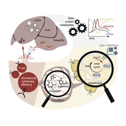
Researchers from the CeMM Research Center for Molecular Medicine of the Austrian Academy of Sciences in collaboration with the Vetmeduni Vienna (including members of Richard Moriggl’s group), MedUni Vienna, Hannover Medical School, St. Gallen Cantonal Hospital and Bio-Cancer Treatment International Ltd identified a key mechanism that explains how antiviral immune responses can reprogram liver metabolism. 

The liver is a crucial organ for the systemic metabolism in our body. In addition to the turnover of biomolecules and drug metabolism, the liver removes toxic substances from the organism. The liver is thus a central metabolic hub in a healthy organism, but is also a central organ in the immune defence against infections - previous studies have shown how immune cells improve the liver metabolism to fight pathogens or cancer. Based on this, the authors of the article now published in the journal Immunity examined these immune metabolic changes during viral infection of mice. In addition to the expected inflammatory changes, the authors identified strong changes in liver metabolism. They were able to show that many central metabolic pathways, including the urea cycle, are suppressed when an infection occurs. The antiviral cytokine type I interferon (IFN-I) was then identified as a regulator of the urea cycle - after removal of the receptor for IFN-I from the surface of hepatocytes, the metabolic changes were no longer observed. This was a surprising observation that shows that IFN-I influences important biological processes during an infection. The results shed a new light on how the body's immune system has developed to regulate liver metabolism so that it controls T cell responses while reducing collateral tissue damage during infection.

Published in Immunity

Alexander Lercher*, Anannya Bhattacharya*, Alexandra M. Popa, Michael Caldera, Moritz F. Schlapansky, Hatoon Baazim, Benedikt Agerer, Bettina Gürtl, Lindsay Kosack, Peter Májek, Julia S. Brunner, Dijana Vitko, Theresa Pinter, Jakob-Wendelin Genger, Anna Orlova, Natalia Pikor, Daniela Reil, Maria Ozsvár-Kozma, Ulrich Kalinke, Burkhard Ludewig, Richard Moriggl, Keiryn L Bennett, Jörg Menche, Paul N. Cheng, Gernot Schabbauer, Michael Trauner, Kristaps Klavins and Andreas Bergthaler (*shared first authors)

Doi: https://doi.org/10.1016/j.immuni.2019.10.014

Acute lymphoblastic leukemia (ALL) is a rare form of cancer that commonly affects children, mostly under the age of five years. In the search for new therapeutic options, researchers at Vetmeduni Vienna funded by the FWF SFB ‘JAK-STAT & Chromatin Landscapes’ have discovered cyclin-dependent kinase 8 (CDK8) as part of the disease process and have developed a novel drug treatment line that is pioneering for future cancer therapies.


Using leukemia mouse models, first author Ingeborg Menzl from the Institute of Pharmacology and Toxicology at Vetmeduni Vienna and her colleagues demonstrated that CDK8-deficient leukemia cells show an increase in cell death. Notably, this function of CDK8 in ALL is independent of enzymatic activity, which means that conventional kinase inhibitors are ineffective. The search for CDK8 interaction partners revealed a previously unknown link to mTOR signaling in cancer cells.

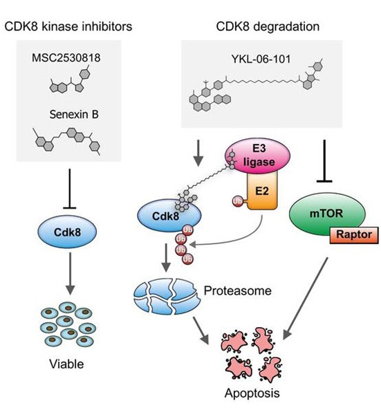
Dual degrader – a therapy line with combined effect. In collaboration with the research team of Nathanael Gray from the Harvard Medical School, the researchers used a new generation of drugs that do not block enzymatic activity but induce the degradation of proteins (called PROTACs). Using a newly synthesized PROTAC mTOR signaling was blocked while simultaneously CDK8 was degraded. With this concept of a dual degrader, the researchers are pioneering for future cancer therapies.


Publication in Nature Communications

Ingeborg Menzl, Tinghu Zhang, Angelika Berger-Becvar, Reinhard Grausenburger, Gerwin Heller, Michaela Prchal-Murphy, Leo Edlinger, Vanessa M. Knab, Iris Z. Uras, Eva Grundschober, Karin Bauer, Mareike Roth, Anna Skucha, Yao Liu, John M. Hatcher, Yanke Liang, Nicholas P. Kwiatkowski, Daniela Fux, Andrea Hoelbl-Kovacic, Stefan Kubicek, Junia V. Melo, Peter Valent, Thomas Weichhart, Florian Grebien, Johannes Zuber, Nathanael S. Gray and Veronika Sexl;

Doi: https://doi.org/10.1038/s41467-019-12656-x

Concept of a Dual Degrader, © Ingeborg Menzl

Acute lymphoblastic leukemia (ALL) is a rare cancer that affects mostly affects children. In the search for new therapeutic options, researchers at Vetmeduni Vienna have now discovered a new mechanism of disease development and proposed a completely new treatment - a pioneering work for future cancer therapies. The study has just been published in Nature Communications.


Cyclin-dependent kinases (CDKs) are frequently deregulated in cancer and represent promising drug targets. The research team of Veronika Sexl at the Vetmeduni Vienna - in collaboration with the research team of Nathanael Gray from Harvard Medical School (USA) - focused on CDK8 in the search for new therapeutic routes for ALL. The reason for this is that tumorigenic cells are dependent on CDK8 function, while healthy cells are not. This opens up a therapeutic window by targeting CDK8: healthy cells are spared while cancer cells will be affected.

The research team was able to show that leukemia cells that lose CDK8 in leukemia mouse models significantly enhance disease latency and prevents disease maintenance. Furthermore, CDK8-depleted cancer cells are highly sensitive to mTOR inhibitors, a previously unknown connection. Thus, the authors have synthesized a small molecule (YKL-06-101) that combines mTOR inhibition and degradation of CDK8, and induces cell death in human leukemic cells. This represents a new therapeutic line in drug development: a dual degrader drug is sufficient to break down a molecule - CDK8 - and at the same time enzymatically block a signalling pathway. They propose that by affecting both simultaneously a potential therapeutic strategy for the treatment of ALL patients might be developed.

Published in Nature Communications

Ingeborg Menzl, Tinghu Zhang, Angelika Berger-Becvar, Reinhard Grausenburger, Gerwin Heller, Michaela Prchal-Murphy, Leo Edlinger, Vanessa M. Knab, Iris Z. Uras, Eva Grundschober, Karin Bauer, Mareike Roth, Anna Skucha, Yao Liu, John M. Hatcher, Yanke Liang, Nicholas P. Kwiatkowski, Daniela Fux, Andrea Hoelbl-Kovacic, Stefan Kubicek, Junia V. Melo, Peter Valent , Thomas Weichhart, Florian Grebien, Johannes Zuber, Nathanael S. Gray and Veronika Sexl

Doi: https://doi.org/10.1038/s41467-019-12656-x

Iris Uras Jodl, from the Institute of Pharmacology and Toxicology at Vetmeduni Vienna, was awarded the Wilhelm Türk Prize of the Austrian Society for Hematology & Medical Oncology for the best scientific work in the field of hematology. The award ceremony took place on October 12th 2019 as part of the joint annual conference of the German, Austrian and Swiss societies for hematology and medical oncology, in Berlin.

The Wilhelm Türk Prize is primarily intended to promote young scientists and is awarded once a year for outstanding research work in the field of hematology. The award is endowed with € 5,000. This year Iris Uras was able to convince the reviewers with her article "Cdk6 coordinates Jak2 V617F mutant MPN via NFkB and apoptotic networks" published in journal Blood, under the supervision of Veronika Sexl. Uras Jodl researched the role of Cyclin-dependent kinase 6 (CDK6) in the development of myeloproliferative neoplasia (MPN) using mouse models.

Veronika Sexl, Head of the Institute for Pharmacology and Toxicology at Vetmeduni Vienna, and Florian Grebien, Head of the Institute for Medical Biochemistry at Vetmeduni Vienna, are working on survival rates of leukemia patients. They are financially supported by the European Research Council (ERC). They have talked about teamwork, crazy concepts and their intersections with veterinary medicine in the VETMED conversation.

The protein CDK6 is at the centre of Veronika Sexl's scientific work at the Institute of Pharmacology and Toxicology. "Starting with this tiny molecule, we're doing broad research," explains the scientist. It has long been known that CDK6 plays a role in many cancers because it drives cell division. It is particularly present in higher amounts in the case of leukemia. "This is why many cancer researchers are working on targeting this protein in the cells by developing special inhibitors against CDK6", says Veronika Sexl. "For the first time we were able to show that CDK6 can and does much more than previously thought."

With the same general goal, but with a different research approach, Florian Grebien is also interested in developing new treatments for cancer. His focus is on the molecular mechanisms behind the development of leukemia. “Our focus is on the so-called fusion proteins”, says Florian Grebien. “They arise when chromosomes break and are incorrectly reassembled. Cancer is very often driven by these fusion proteins. If you study them functionally, you can learn a lot about what processes a cell needs to become a cancer cell”.

Both researchers and their projects are currently funded by European Research Council (ERC) grants. In 2016 Sexl received an “Advanced Grant” worth 2.5 million euros. Florian Grebien was awarded a “Starting Grant” worth 1.5 million euros in 2015.

The full interview can be found here.

Langerhans cell histiocytosis (LCH) is a rare disease that mainly affects small children. It occupies a hybrid position between cancers and inflammatory diseases, which makes it an attractive model for studying cancer development. While LCH can heal itself in some patients, other cases require intensive chemotherapy with long-term consequences for the children. The reasons for these differences are hardly known. In a new study published in the journal Cancer Discovery, researchers from St. Anna Children's Cancer Research Institute (CCRI) and the Center for Molecular Medicine of the Austrian Academy of Sciences (CeMM) uncovered important insights into the cellular heterogeneity and molecular mechanisms of LCH.

Caroline Hutter, pediatric oncologist at St. Anna Children's Hospital, observed a remarkable heterogeneity between LCH cells when examining LCH lesions under the microscope. In order to investigate this diversity in detail, she assembled an interdisciplinary team of experimental and computer researchers from CCRI and CeMM, as well as physicians from St. Anna Children's Hospital and General Hospital in Vienna. Caroline Hutter's aim is to answer two fundamental questions: What are the mechanisms behind LCH and how can we improve the treatment of children affected by this disease?

LCH lesions were analysed in the laboratory of Christoph Bock (CeMM), by Florian Halbritter (now at CCRI) and Matthias Farlik (now at the MedUni Vienna), with sufficient resolution to identify the molecular patterns of individual cells in detail and to develop a comprehensive "map" of cellular heterogeneity in LCH. On this molecular map, the team identified several subtypes of LCH cells. Among them was a group of actively dividing cells that are believed to be the precursors of other LCH cells. The team deciphered the molecular signalling pathways that are active in different branches of this unexpected developmental hierarchy, highlighting an interplay of developmental, immunological and oncogenic mechanisms in LCH. In the future, these findings could help to better differentiate between severe and less severe cases of the disease and even open up new treatment options.

Published in Cancer Discovery
Florian Halbritter, Matthias Farlik, Raphaela Schwentner, Gunhild Jug, Nikolaus Fortelny,
Thomas Schnoller, Hanja Pisa, LindaChristina Schuster, Andrea Reinprecht, Thomas Czech,
Johannes Gojo, Wolfgang Holter, Milen Minkov, Wolfgang M Bauer, Ingrid Simonitsch Klupp, Christoph Bock and Caroline Hutter

Doi:10.1158/2159-8290.CD-19-0138

New insights into the development of an unusual childhood disease

The interferons initiate a signaling process that causes the cell to activate the protein complex ISGF3 for driving antimicrobial gene expression. Scientists led by Thomas Decker at the Max Perutz Labs now found out that two of the three proteins forming this complex are permanently present at these genes, independently of the activating cascade caused by interferons. STAT2-IRF9 forms this ‘light’ version of ISGF3 and allows for homeostatic low expression of antimicrobial genes. Upon pathogen recognition interferons are produced and activate the complete version of ISGF3 composed of STAT1-STAT2-IRF9.  This trimeric ISGF3 switches to a full-fledged antimicrobial transcriptional program. The homeostatic presence of STAT2-IRF9 at antimicrobial genes governs cellular alertness and the rapid exchange to the interferon induced complete ISGF3 explains how the innate immune system activates in such a quick manner.


Publication in Nature Communications

Ekaterini Platanitis, Duygu Demiroz, Anja Schneller, Katrin Fischer, Christophe Capelle, Markus Hartl, Thomas Gossenreiter, Mathias Müller, Maria Novatchkova and Thomas Decker

A molecular switch from STAT2-IRF9 to ISGF3 underlies interferon-induced gene transcription (2019);

Doi: https://doi.org/10.1038/s41467-019-10970-y