Wilfried Ellmeier,
Division of Immunobiology,
Institute of Immunology, MedUni Vienna;
Coordinator of newly granted SFB F70 ‘HDACs as regulators of T cell-mediated immunity in health and disease’
(http://www.meduniwien.ac.at/HIT/)

Title: Histone deacetylases and the control of CD4+ T cell-mediated immunity

http://www.meduniwien.ac.at/immunologie/ellmeier

The long-term research interest of Wilfried Ellmeier’s group is to characterize molecular mechanisms that regulate the development and function of T lymphocytes. They aim to provide important and medical relevant insight into the regulation of T cell-mediated immunity.


We used conditional ablation of STAT1 in macrophages, monocytes and granulocytes of mice and showed that myeloid STAT1 protects from early murine cytomegalovirus replication and pathology in spleen. Unexpectedly, we found that myeloid STAT1 drives extramedullary haematopoiesis (EMH). STAT1 promotes EMH not only after virus infection but also upon sterile inflammation induced by CpG oligodeoxynucleotides. Using additional genetically engineered mice we analysed the impact of upstream signals known to activate STAT1 and observed that virus-induced EMH does not rely on type I or type II IFN signaling in myeloid cells or IL-27 signaling in all cell types. Our studies provide the first genetic evidence that STAT1-dependent signaling in myeloid cells restricts MCMV at early time points post infection and induces compensatory (stress-induced) haematopoiesis in the spleen.

Publication in Cell Reports

Riem Gawish*, Tanja Bulat*, Mario Biaggio*, Caroline Lassnig, Zsuzsanna Bago-Horvath, Sabine Macho-Maschler, Andrea Poelzl, Natalija Simonović, Michaela Prchal-Murphy, Rita Rom, Lena Amenitsch, Luca Ferrarese, Juliana Kornhoff, Therese Lederer, Jasmin Svinka, Robert Eferl, Markus Bosmann, Ulrich Kalinke, Dagmar Stoiber, Veronika Sexl, Astrid Krmpotić, Stipan Jonjić, Mathias Müller, and Birgit Strobl

*equal author contribution

Myeloid Cells Restrict MCMV and Drive Stress-Induced Extramedullary Hematopoiesis through STAT1 (2019); Doi: https://doi.org/10.1016/j.celrep.2019.02.017

We used conditional ablation of TYK2 in mice and showed that TYK2 promotes NK cell activity in tumour surveillance and the defence against Listeria monocytogenes infection through cell-extrinsic and -intrinsic mechanisms. The key discoveries are as follows: NK cell-extrinsic TYK2 drives peripheral NK cell maturation, demethylation of the Ifng locus, activating receptor-induced IFNg production, cytotoxicity and anti-tumour activity; the NK cell defects observed in Tyk2-/- mice can be restored by recombinant IL-15/IL-15Rα treatment; NK cell-intrinsic TYK2 signalling mediates infection-induced IFNg production and acts protective during Listeria monocytogenes infection.

Collectively, our study disclosed TYK2 functions that remained unrecognized in mice with complete TYK2 deficiency. Our findings that cytotoxic defects of Tyk2-/- NK cells can be rescued by IL-15/IL-15Rα treatment suggest that unwanted effects of TYK2 inhibitors in tumour therapy may be overcome by boosting NK cell activity.

Publication in Journal of Immunology

Natalija Simonović * , Agnieszka Witalisz-Siepracka *, Katrin Meissl, Caroline Lassnig, Ursula Reichart,  Thomas Kolbe,  Matthias Farlik, Christoph Bock, Veronika Sexl, Mathias Müller, and Birgit Strobl

*equal author contribution

NK Cells Require Cell-Extrinsic and -Intrinsic TYK2 for Full Functionality in Tumor Surveillance and Antibacterial Immunity (2019); Doi:

https://doi.org/10.4049/jimmunol.1701649

The symposium will be held at the Vetmeduni Vienna on January 31 and February 01, 2019. The symposium invited speakers from the Vienna Life Science community and from the Vetmeduni Vienna who work in basic and translational biomedical sciences to pave the road for new therapeutic concepts. Their research contributions serve as role models for students and early career scientists. We train students of the MSc program ‘Comparative Biomedicine’on our campus under the principle of “One Health – One Medicine”. The symposium is organized and supported by SFB F61 JakStat Monarchies.

Link to PDF of Program

An international research team led by researchers from Vetmeduni Vienna have made an important discovery that could lead to a better understanding of lymphocytic leukemia. They identified the STAT5B protein as crucial for the development of the disease. The findings represent a possible therapeutic approach involving new, precision medicine strategies.

The BCR/ABL fusion gene, which does not occur among healthy people, has been shown to be a causative agent in the pathogenesis of B-cell acute lymphocytic leukemia (ALL). This gene leads to transformation of white blood cells, which can proliferate out of control. Earlier studies by the research group of Veronika Sexl at Vetmeduni Vienna showed that the STAT5 transcription factor was essential for the development of BCR/ABL-induced leukemia. Surprisingly, mutations in StatB, but not StatA, have been frequently described in hematopoietic tumors; therefore, the authors of this new study used BCR/ABL as a model system to disentangle the contribution of STAT5A or STAT5B for leukemogenesis. They found that the absence of STAT5A led to a decrease in cell survival and the formation of colonies of malignant cancer cells; the effects were even more drastic in the absence of STAT5B. In the mouse model, loss of STAT5B increased interferon response and suppressed transformation. The opposite scenario was true in patients with overactive STAT5B: the interferon response against tumour growth was suppressed and transformation was enhanced. According to the researchers, this may be of direct clinical relevance for patients, as a better understanding of the complex role of STAT5B could enable the development of precision medicine strategies to treat BCR/ABL ALL.

Published in Leukemia

Sebastian Kollmann, Eva Grundschober, Barbara Maurer, Wolfgang Warsch, Reinhard Grausenburger, Leo Edlinger, Jani Huuhtanen, Sabine Lagger, Lothar Hennighausen, Peter Valente, Thomas Decker, Birgit Strobl, Mathias Müller, Satu Mustjoki, Andrea Hölbl-Kovacic and Veronika Sexl.

Doi: https://doi.org/10.1038/s41375-018-0369-5

Robert Eferl,
Institute of Cancer Research,
MedUni Vienna

Title: CDHR5 in intestinal tissue homeostasis and cancer

https://krebsforschung.meduniwien.ac.at/forschung-research/research-focuses/cellular-and-molecular-tumor-biology/robert-eferl/

Robert Eferl works in two main topics:
1) identification of cooperative signaling pathways in cancer formation using novel mouse models.
2) functions of STAT transcription factors in inflammatory liver damage and cancer types that are associated with inflammation

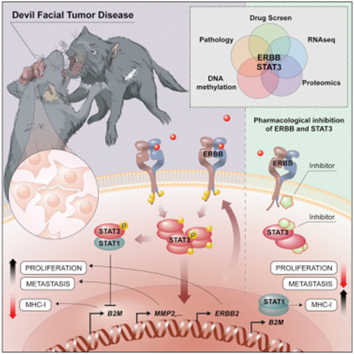
Facial tumours of Tasmanian devils belong to rare cases of transmissible cancers. More than 90% of the population of devils is extinct due to two distinct Schwannoma cell lines that spread among the species. They are of great interest for biomedical research, as they allow the study of fundamental properties of cancer cells and how they escape the host´s immune system. Using cutting-edge technologies, scientists of the group of Andreas Bergthaler at the CeMM Research Center for Molecular Medicine of the Austrian Academy of Sciences and members of the groups of Richard Moriggl and Christoph Bock, both SFB JakStat Monarchies at the Vienna University of Veterinary Medicine together with international collaborators identified the ERBB receptor-STAT3 axis as key molecular mechanisms allowing for the transmissibility of the tumour cells. Importantly, the consortium showed that the inhibition of ERBB receptors with a selective tyrosine kinase inhibitor or pharmacologic intervention of STAT3 could selectively kill the transmissible cancer cells upregulating MHC class I allowing for immune cell recognition. This could play an important role for the treatment and understanding of the disease to rescue the Tasmanian devil in the isolated island from extinction.

Publication in Cancer Cell:

Lindsay Kosack, Bettina Wingelhofer, Alexandra Popa, Anna Orlova, Benedikt Agerer, Bojan Vilagos, Peter Majek, Katja Parapatics, Alexander Lercher, Anna Ringler, Johanna Klughammer, Mark Smyth, Kseniya Khamina, Hatoon Baazim, Elvin D. de Araujo, David A. Rosa, Jisung Park, Gary Tin, Siawash Ahmar, Patrick T. Gunning, Christoph Bock, Hannah V. Siddle, Gregory M. Woods, Stefan Kubicek, Elisabeth P. Murchison, Keiryn L. Bennett, Richard Moriggl and Andreas Bergthaler

The ERBB-STAT3 Axis Drives Tasmanian Devil Facial Tumor Disease (2019) DOI:https://doi.org/10.1016/j.ccell.2018.11.018

See also “The Deadly Bite of STAT3” mini review Cancer Cell by Hagen Schwenzer and Ariberto Fassati: DOI:https://doi.org/10.1016/j.ccell.2018.12.004

Facial tumours of Tasmanian Devils are among the extremely rare cases of transmissible cancer and threaten the survival of this species. They are extremely interesting for medical research because they can be used to unravel new cancer molecular mechanisms and their interplay with the immune system. Scientists at the Center for Molecular Medicine (CeMM), the Vetmeduni Vienna and the MedUni Vienna have succeeded in using modern analysis methods to elucidate key molecular mechanisms that are crucial for the transferability of these cancer cells.

Tumours can usually only proliferate in the organism from which they originated. This is due to a large number of molecular security mechanisms with which the immune system repels and destroys foreign tissue. An exception to this is the Tasmanian Devil, the largest living carnivorous marsupial in the world: a deadly facial tumour has been spreading at a rapid pace for over two decades in this species.

How these cells escape the immune system of its recipient has long been a mystery. Now, the groups of Andreas Bergthaler (CeMM) and Richard Moriggl, head of the Ludwig Boltzmann Institute for Cancer Research and Professor of Functional Cancer Genomics at the Vetmeduni and MedUni Vienna,

found that certain receptor molecules on the surface of the cancer cells, ERBB receptors, have unusually high activity. The activation of these receptors influences the cell's epigenetic make-up via STAT3. As a result, the number of molecules that the immune system uses to recognize the cell is reduced, while cell division accelerates and factors for the metastasis of the tumour cells are increasingly produced. Furthermore, inhibiting the ERBB receptor with a specific drug can kill cancer cells in a targeted manner. This could play an important role in treating this communicable tumour before the Tasmanian Devil is completely eradicated. In addition, the basic biological principles of invasion and fixation in new tissues are of crucial importance, even in the case of non-communicable tumours, in particular cancer metastases. A better molecular understanding of these relatively exotic communicable tumours can provide important insights into the basic biological mechanisms of cancer.

Published in Cancer Cell

Lindsay Kosack, Bettina Wingelhofer, Alexandra Popa, Anna Orlova, Benedikt Agerer, Bojan Vilagos, Peter Majek, Katja Parapatics, Alexander Lercher, Anna Ringler, Johanna Klughammer, Mark Smyth, Kseniya Khamina, Hatoon Baazim, Elvin D. de Araujo, David A. Rosa, Jisung Park, Gary Tin, Siawash Ahmar, Patrick T. Gunning, Christoph Bock, Hannah V. Siddle, Gregory M. Woods, Stefan Kubicek, Elisabeth P. Murchison, Keiryn L. Bennett, Richard Moriggl and Andreas Bergthaler

Doi: https://doi.org/10.1016/j.ccell.2018.11.018