Acute lymphoblastic leukemia (ALL) is a rare form of cancer that commonly affects children, mostly under the age of five years. In the search for new therapeutic options, researchers at Vetmeduni Vienna funded by the FWF SFB ‘JAK-STAT & Chromatin Landscapes’ have discovered cyclin-dependent kinase 8 (CDK8) as part of the disease process and have developed a novel drug treatment line that is pioneering for future cancer therapies.


Using leukemia mouse models, first author Ingeborg Menzl from the Institute of Pharmacology and Toxicology at Vetmeduni Vienna and her colleagues demonstrated that CDK8-deficient leukemia cells show an increase in cell death. Notably, this function of CDK8 in ALL is independent of enzymatic activity, which means that conventional kinase inhibitors are ineffective. The search for CDK8 interaction partners revealed a previously unknown link to mTOR signaling in cancer cells.

Dual degrader – a therapy line with combined effect. In collaboration with the research team of Nathanael Gray from the Harvard Medical School, the researchers used a new generation of drugs that do not block enzymatic activity but induce the degradation of proteins (called PROTACs). Using a newly synthesized PROTAC mTOR signaling was blocked while simultaneously CDK8 was degraded. With this concept of a dual degrader, the researchers are pioneering for future cancer therapies.


Publication in Nature Communications

Ingeborg Menzl, Tinghu Zhang, Angelika Berger-Becvar, Reinhard Grausenburger, Gerwin Heller, Michaela Prchal-Murphy, Leo Edlinger, Vanessa M. Knab, Iris Z. Uras, Eva Grundschober, Karin Bauer, Mareike Roth, Anna Skucha, Yao Liu, John M. Hatcher, Yanke Liang, Nicholas P. Kwiatkowski, Daniela Fux, Andrea Hoelbl-Kovacic, Stefan Kubicek, Junia V. Melo, Peter Valent, Thomas Weichhart, Florian Grebien, Johannes Zuber, Nathanael S. Gray and Veronika Sexl;

Doi: https://doi.org/10.1038/s41467-019-12656-x

Concept of a Dual Degrader, © Ingeborg Menzl

Acute lymphoblastic leukemia (ALL) is a rare cancer that affects mostly affects children. In the search for new therapeutic options, researchers at Vetmeduni Vienna have now discovered a new mechanism of disease development and proposed a completely new treatment - a pioneering work for future cancer therapies. The study has just been published in Nature Communications.


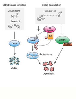
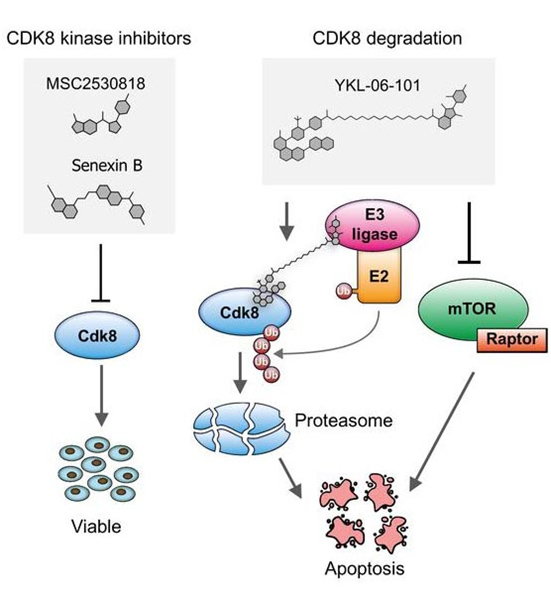
Cyclin-dependent kinases (CDKs) are frequently deregulated in cancer and represent promising drug targets. The research team of Veronika Sexl at the Vetmeduni Vienna - in collaboration with the research team of Nathanael Gray from Harvard Medical School (USA) - focused on CDK8 in the search for new therapeutic routes for ALL. The reason for this is that tumorigenic cells are dependent on CDK8 function, while healthy cells are not. This opens up a therapeutic window by targeting CDK8: healthy cells are spared while cancer cells will be affected.

The research team was able to show that leukemia cells that lose CDK8 in leukemia mouse models significantly enhance disease latency and prevents disease maintenance. Furthermore, CDK8-depleted cancer cells are highly sensitive to mTOR inhibitors, a previously unknown connection. Thus, the authors have synthesized a small molecule (YKL-06-101) that combines mTOR inhibition and degradation of CDK8, and induces cell death in human leukemic cells. This represents a new therapeutic line in drug development: a dual degrader drug is sufficient to break down a molecule - CDK8 - and at the same time enzymatically block a signalling pathway. They propose that by affecting both simultaneously a potential therapeutic strategy for the treatment of ALL patients might be developed.

Published in Nature Communications

Ingeborg Menzl, Tinghu Zhang, Angelika Berger-Becvar, Reinhard Grausenburger, Gerwin Heller, Michaela Prchal-Murphy, Leo Edlinger, Vanessa M. Knab, Iris Z. Uras, Eva Grundschober, Karin Bauer, Mareike Roth, Anna Skucha, Yao Liu, John M. Hatcher, Yanke Liang, Nicholas P. Kwiatkowski, Daniela Fux, Andrea Hoelbl-Kovacic, Stefan Kubicek, Junia V. Melo, Peter Valent , Thomas Weichhart, Florian Grebien, Johannes Zuber, Nathanael S. Gray and Veronika Sexl

Doi: https://doi.org/10.1038/s41467-019-12656-x

Langerhans cell histiocytosis (LCH) is a rare disease that mainly affects small children. It occupies a hybrid position between cancers and inflammatory diseases, which makes it an attractive model for studying cancer development. While LCH can heal itself in some patients, other cases require intensive chemotherapy with long-term consequences for the children. The reasons for these differences are hardly known. In a new study published in the journal Cancer Discovery, researchers from St. Anna Children's Cancer Research Institute (CCRI) and the Center for Molecular Medicine of the Austrian Academy of Sciences (CeMM) uncovered important insights into the cellular heterogeneity and molecular mechanisms of LCH.

Caroline Hutter, pediatric oncologist at St. Anna Children's Hospital, observed a remarkable heterogeneity between LCH cells when examining LCH lesions under the microscope. In order to investigate this diversity in detail, she assembled an interdisciplinary team of experimental and computer researchers from CCRI and CeMM, as well as physicians from St. Anna Children's Hospital and General Hospital in Vienna. Caroline Hutter's aim is to answer two fundamental questions: What are the mechanisms behind LCH and how can we improve the treatment of children affected by this disease?

LCH lesions were analysed in the laboratory of Christoph Bock (CeMM), by Florian Halbritter (now at CCRI) and Matthias Farlik (now at the MedUni Vienna), with sufficient resolution to identify the molecular patterns of individual cells in detail and to develop a comprehensive "map" of cellular heterogeneity in LCH. On this molecular map, the team identified several subtypes of LCH cells. Among them was a group of actively dividing cells that are believed to be the precursors of other LCH cells. The team deciphered the molecular signalling pathways that are active in different branches of this unexpected developmental hierarchy, highlighting an interplay of developmental, immunological and oncogenic mechanisms in LCH. In the future, these findings could help to better differentiate between severe and less severe cases of the disease and even open up new treatment options.

Published in Cancer Discovery
Florian Halbritter, Matthias Farlik, Raphaela Schwentner, Gunhild Jug, Nikolaus Fortelny,
Thomas Schnoller, Hanja Pisa, LindaChristina Schuster, Andrea Reinprecht, Thomas Czech,
Johannes Gojo, Wolfgang Holter, Milen Minkov, Wolfgang M Bauer, Ingrid Simonitsch Klupp, Christoph Bock and Caroline Hutter

Doi:10.1158/2159-8290.CD-19-0138

New insights into the development of an unusual childhood disease

The interferons initiate a signaling process that causes the cell to activate the protein complex ISGF3 for driving antimicrobial gene expression. Scientists led by Thomas Decker at the Max Perutz Labs now found out that two of the three proteins forming this complex are permanently present at these genes, independently of the activating cascade caused by interferons. STAT2-IRF9 forms this ‘light’ version of ISGF3 and allows for homeostatic low expression of antimicrobial genes. Upon pathogen recognition interferons are produced and activate the complete version of ISGF3 composed of STAT1-STAT2-IRF9.  This trimeric ISGF3 switches to a full-fledged antimicrobial transcriptional program. The homeostatic presence of STAT2-IRF9 at antimicrobial genes governs cellular alertness and the rapid exchange to the interferon induced complete ISGF3 explains how the innate immune system activates in such a quick manner.


Publication in Nature Communications

Ekaterini Platanitis, Duygu Demiroz, Anja Schneller, Katrin Fischer, Christophe Capelle, Markus Hartl, Thomas Gossenreiter, Mathias Müller, Maria Novatchkova and Thomas Decker

A molecular switch from STAT2-IRF9 to ISGF3 underlies interferon-induced gene transcription (2019);

Doi: https://doi.org/10.1038/s41467-019-10970-y

In a triple-effort between international research groups from the University of Veterinary Medicine Vienna, Harvard University and the University of Toronto, important new information was discovered about the protein STAT5B, which is mutated in patients with T-cell cancers. STAT5B, like all proteins, is made up of building blocks called amino acids. A single amino acid change in STAT5B makes it hyperactive and leads to T-cell cancer development. We have tackled the difficult task to visualize the structure and shape of STAT5B in order to facilitate the discovery of new drugs that specifically target the mutant cancer-causing form of the protein, whilst sparing the important normal-functioning STAT5B.

We have used a technique similar to medical X-rays to reveal for the first time the three-dimensional structures of normal and mutant STAT5B down to the atomic level. We also developed a new cancer mouse model driven by mutant STAT5B, which allows the study of one of the most aggressive T-cell cancers seen in patients. Importantly, the structural information and the disease model can now be used to test new drugs that target only the cancer-causing form of STAT5B, which will significantly reduce the side-effects and increase the effectiveness of the treatment.

Publication in Nature Communications

Elvin D. de Araujo*, Fettah Erdogan*, Heidi A. Neubauer*, Deniz Meneksedag-Erol, Pimyupa Manaswiyoungkul, Mohammad S. Eram, Hyuk-Soo Seo, Abdul K. Qadree, Johan Israelian, Anna Orlova, Tobias Suske, Ha T. T. Pham, Auke Boersma, Simone Tangermann, Lukas Kenner, Thomas Rülicke, Aiping Dong, Manimekalai Ravichandran, Peter J. Brown, Gerald F. Audette, Sarah Rauscher, Sirano Dhe-Paganon, Richard Moriggl and Patrick T. Gunning

*equal author contribution; corresponding authorship

Structural and functional consequences of the STAT5BN642H driver mutation (2019); Doi: https://doi.org/10.1038/s41467-019-10422-7

An international research team led by Veronika Sexl from Vetmeduni Vienna and supported by other members of SFB ‚JakStat Monarchies‘ have made an important discovery that could lead to a better understanding of lymphocytic leukemia: the STAT5B protein – but not the highly related STAT5A isoform – is crucial for disease development by suppressing interferon signals during leukemic transformation. Our findings will enable novel therapeutic approaches in precision medicine.

Publication in Leukemia:

Sebastian Kollmann, Eva Grundschober, Barbara Maurer, Wolfgang Warsch, Reinhard Grausenburger, Leo Edlinger, Jani Huuhtanen, Sabine Lagger, Lothar Hennighausen, Peter Valent, Thomas Decker, Birgit Strobl, Mathias Mueller, Satu Mustjoki, Andrea Hoelbl-Kovacic and Veronika Sexl

Twins with different personalities: STAT5B—but not STAT5A—has a key role in BCR/ABL-induced leukemia (2019), https://doi.org/10.1038/s41375-018-0369-5

We used conditional ablation of STAT1 in macrophages, monocytes and granulocytes of mice and showed that myeloid STAT1 protects from early murine cytomegalovirus replication and pathology in spleen. Unexpectedly, we found that myeloid STAT1 drives extramedullary haematopoiesis (EMH). STAT1 promotes EMH not only after virus infection but also upon sterile inflammation induced by CpG oligodeoxynucleotides. Using additional genetically engineered mice we analysed the impact of upstream signals known to activate STAT1 and observed that virus-induced EMH does not rely on type I or type II IFN signaling in myeloid cells or IL-27 signaling in all cell types. Our studies provide the first genetic evidence that STAT1-dependent signaling in myeloid cells restricts MCMV at early time points post infection and induces compensatory (stress-induced) haematopoiesis in the spleen.

Publication in Cell Reports

Riem Gawish*, Tanja Bulat*, Mario Biaggio*, Caroline Lassnig, Zsuzsanna Bago-Horvath, Sabine Macho-Maschler, Andrea Poelzl, Natalija Simonović, Michaela Prchal-Murphy, Rita Rom, Lena Amenitsch, Luca Ferrarese, Juliana Kornhoff, Therese Lederer, Jasmin Svinka, Robert Eferl, Markus Bosmann, Ulrich Kalinke, Dagmar Stoiber, Veronika Sexl, Astrid Krmpotić, Stipan Jonjić, Mathias Müller, and Birgit Strobl

*equal author contribution

Myeloid Cells Restrict MCMV and Drive Stress-Induced Extramedullary Hematopoiesis through STAT1 (2019); Doi: https://doi.org/10.1016/j.celrep.2019.02.017

We used conditional ablation of TYK2 in mice and showed that TYK2 promotes NK cell activity in tumour surveillance and the defence against Listeria monocytogenes infection through cell-extrinsic and -intrinsic mechanisms. The key discoveries are as follows: NK cell-extrinsic TYK2 drives peripheral NK cell maturation, demethylation of the Ifng locus, activating receptor-induced IFNg production, cytotoxicity and anti-tumour activity; the NK cell defects observed in Tyk2-/- mice can be restored by recombinant IL-15/IL-15Rα treatment; NK cell-intrinsic TYK2 signalling mediates infection-induced IFNg production and acts protective during Listeria monocytogenes infection.

Collectively, our study disclosed TYK2 functions that remained unrecognized in mice with complete TYK2 deficiency. Our findings that cytotoxic defects of Tyk2-/- NK cells can be rescued by IL-15/IL-15Rα treatment suggest that unwanted effects of TYK2 inhibitors in tumour therapy may be overcome by boosting NK cell activity.

Publication in Journal of Immunology

Natalija Simonović * , Agnieszka Witalisz-Siepracka *, Katrin Meissl, Caroline Lassnig, Ursula Reichart,  Thomas Kolbe,  Matthias Farlik, Christoph Bock, Veronika Sexl, Mathias Müller, and Birgit Strobl

*equal author contribution

NK Cells Require Cell-Extrinsic and -Intrinsic TYK2 for Full Functionality in Tumor Surveillance and Antibacterial Immunity (2019); Doi:

https://doi.org/10.4049/jimmunol.1701649

An international research team led by researchers from Vetmeduni Vienna have made an important discovery that could lead to a better understanding of lymphocytic leukemia. They identified the STAT5B protein as crucial for the development of the disease. The findings represent a possible therapeutic approach involving new, precision medicine strategies.

The BCR/ABL fusion gene, which does not occur among healthy people, has been shown to be a causative agent in the pathogenesis of B-cell acute lymphocytic leukemia (ALL). This gene leads to transformation of white blood cells, which can proliferate out of control. Earlier studies by the research group of Veronika Sexl at Vetmeduni Vienna showed that the STAT5 transcription factor was essential for the development of BCR/ABL-induced leukemia. Surprisingly, mutations in StatB, but not StatA, have been frequently described in hematopoietic tumors; therefore, the authors of this new study used BCR/ABL as a model system to disentangle the contribution of STAT5A or STAT5B for leukemogenesis. They found that the absence of STAT5A led to a decrease in cell survival and the formation of colonies of malignant cancer cells; the effects were even more drastic in the absence of STAT5B. In the mouse model, loss of STAT5B increased interferon response and suppressed transformation. The opposite scenario was true in patients with overactive STAT5B: the interferon response against tumour growth was suppressed and transformation was enhanced. According to the researchers, this may be of direct clinical relevance for patients, as a better understanding of the complex role of STAT5B could enable the development of precision medicine strategies to treat BCR/ABL ALL.

Published in Leukemia

Sebastian Kollmann, Eva Grundschober, Barbara Maurer, Wolfgang Warsch, Reinhard Grausenburger, Leo Edlinger, Jani Huuhtanen, Sabine Lagger, Lothar Hennighausen, Peter Valente, Thomas Decker, Birgit Strobl, Mathias Müller, Satu Mustjoki, Andrea Hölbl-Kovacic and Veronika Sexl.

Doi: https://doi.org/10.1038/s41375-018-0369-5

Facial tumours of Tasmanian devils belong to rare cases of transmissible cancers. More than 90% of the population of devils is extinct due to two distinct Schwannoma cell lines that spread among the species. They are of great interest for biomedical research, as they allow the study of fundamental properties of cancer cells and how they escape the host´s immune system. Using cutting-edge technologies, scientists of the group of Andreas Bergthaler at the CeMM Research Center for Molecular Medicine of the Austrian Academy of Sciences and members of the groups of Richard Moriggl and Christoph Bock, both SFB JakStat Monarchies at the Vienna University of Veterinary Medicine together with international collaborators identified the ERBB receptor-STAT3 axis as key molecular mechanisms allowing for the transmissibility of the tumour cells. Importantly, the consortium showed that the inhibition of ERBB receptors with a selective tyrosine kinase inhibitor or pharmacologic intervention of STAT3 could selectively kill the transmissible cancer cells upregulating MHC class I allowing for immune cell recognition. This could play an important role for the treatment and understanding of the disease to rescue the Tasmanian devil in the isolated island from extinction.

Publication in Cancer Cell:

Lindsay Kosack, Bettina Wingelhofer, Alexandra Popa, Anna Orlova, Benedikt Agerer, Bojan Vilagos, Peter Majek, Katja Parapatics, Alexander Lercher, Anna Ringler, Johanna Klughammer, Mark Smyth, Kseniya Khamina, Hatoon Baazim, Elvin D. de Araujo, David A. Rosa, Jisung Park, Gary Tin, Siawash Ahmar, Patrick T. Gunning, Christoph Bock, Hannah V. Siddle, Gregory M. Woods, Stefan Kubicek, Elisabeth P. Murchison, Keiryn L. Bennett, Richard Moriggl and Andreas Bergthaler

The ERBB-STAT3 Axis Drives Tasmanian Devil Facial Tumor Disease (2019) DOI:https://doi.org/10.1016/j.ccell.2018.11.018

See also “The Deadly Bite of STAT3” mini review Cancer Cell by Hagen Schwenzer and Ariberto Fassati: DOI:https://doi.org/10.1016/j.ccell.2018.12.004